VisionSlit — Computer Vision Integrated Slit Lamp
High-resolution slit-lamp capture with real-time AI to flag cataract, glaucoma and corneal disease; generates structured, image-rich reports and supports longitudinal comparisons.
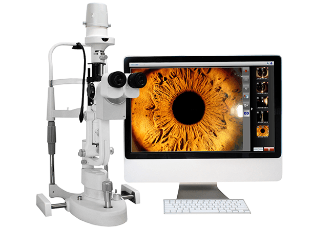
Overview
VisionSlit is a next-generation ophthalmic diagnostic tool that integrates a high-resolution camera and real-time computer vision analysis into the slit-lamp exam. As the clinician performs the exam, images and short videos are captured and analyzed to flag features consistent with cataract, glaucoma, and corneal pathology. The system generates structured, image-rich reports and can retrieve prior visits for side-by-side comparison, supporting continuity and objective monitoring over time.
Clinical Problem
Traditional slit-lamp documentation often relies on free-text notes and memory, which vary between clinicians and across visits. Subtle early signs may be missed, and longitudinal comparisons are difficult without standardized images and formats. Clinics need a way to standardize capture, provide automated analytical hints, and produce reusable, EMR-ready outputs that can be compared over months or years.
Methodology
- Embed a high-resolution ophthalmic camera to capture stills/video during routine slit-lamp examination.
- Apply computer-vision models for lesion/anomaly detection and present annotated findings in real time.
- Store cases with standardized metadata and enable past-vs-present side-by-side comparisons in the report.
Tech Stack
Equipment
Expected Outcomes
Prototype prepared for initial clinical trials with ophthalmology partners.